Glossary
The following are terms to know in alphabetical order (not in an order that you should learn these concepts)
Action Potential
The firing of a neuron, the method that neurons use to transmit data.
Phases
- Resting Phase
- Membrane at resting potential
- Stimulation
- something makes the membrane more positive until threshold is hit
- Rising Phase
- Voltage Gated Sodium Channels open.
- Sodium Rushes in, making the membrane potential more positive (depolarization)
- Voltage Gated Sodium Channels open.
- Falling Phase
- Voltage Gated Sodium Channels become inactive, sodium no longer allowed through
- The slower Voltage Gated Potassium channels open
- potassium rushes out of cell, making it more negative
- Undershoot
- Overshoot before voltage gated Potassium channels close
- Recovery
- Gradual return to resting potential
The speed of an action potential is affected by membrane thickness (thicker means higher capacitance) and axon diameter (higher means lower resistance). The fastest speed needs the lowest possible capacitance and resistance.
Capacitance
The storing of electrical charge, which slows down the change of voltage. Measured in Farads (F).
- In neurons, Capacitance is increased by decreasing axon diameter and increasing membrane thickness.
- Decreasing capacitance makes voltage changes faster across the membrane.
- Time constant tau is how long it takes a piece of membrane to charge up to 63% of its final value in response to a step change in its input voltage
- If the dielectric has a higher dielectric constant (how easy it is to polarize the material), the capacitance increases.
- if we increase the distance between the place by making the dielectric thicker, capacitance decreases
- Cells use the fatty lipid bilayer as their dielectric
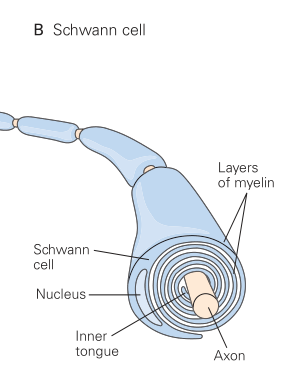
- if cells need to lower capacitance, they will increase their dielectric thickness by wrapping layers around a membrane in a process known as myelination.
Central Nervous System (CNS)
A division of the nervous system that is comprised of the Brain and Spinal Cord.
CerebroSpinal Fluid (CSF)
A fluid that bathes the brain and spinal cord. It acts as a shock absorber and caries nutrients to nervous system cells and waste away from them. It is renewed by the body by volume about 4-5 times a day. It is separated from other bodily fluids by the Blood Brain Barrier (BBB).
Current
The 'amount' of charge flow, measured in Amps (A). Better described as the change of charge over time -> dC/dt
Electrostatic force
The attraction or repulsion between charged particles, where oppositely charged particles.
Example: Negatively charged particles (Anions) are attracted to positively charged particles (Cations)
Diffusion
The tendency of 2 solutions of different concentrations separated by a permeable membrane, to equalize in concentration as particles from the area of high concentration will diffuse to the area of low concentration until Equilibrium. Resultant of the laws of entropy, and generally takes energy to counteract this effect (or other forces, such as interactions between charged particles).
ElectroChemical Gradient
The result of the forces of diffusion and the forces of Electrostatic force working together
Equilibrium
The state of rest when the net movement of particles passing one way through a membrane equals the particles passing through the other way, such that concentrations stay constant.
Equilibrium DOES NOT mean that particles stopped moving, NOR those it mean that the 2 separated concentrations are EQUAL, since forces such electrostatic forces (even between different ions) can change this equilibrium point away from the point of equal concentration
Glial Cells
While Glial cells develop from the same neural progenitor cells in the embryo and often share many characteristics of neurons, glial cells do not directly get involved in signaling in the sense that they are non-conductive. Glia support various functions of neurons. They may coat the axons of neurons to aid in signal transmission, release / absorb proteins to regulate signaling, or be involved in various process crucial for neural plasticity.
There are 2 kinds of glia cells: Macroglia and Microglia.
Macroglia Cells
These glia cells make up 90% of the glia cells in the human brain. They buffer some of the biochemical process in neurons and help clean up neurotransmitters at synapses. They are the ones you hear most often about and there are several major types:
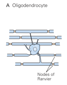
- Oligodendrocytes
- Schwann Cells
- Ependymal Cells
- Involved in the production of CSF
- makes up the Lining of the BBB
- also line the ventricles of the brain
- have apical microvilli to increase surface area for diffusion
- have cilia to promote CSF movement
- capable of cleaning the CSF itself
- Astrocytes
- Star shaped, have lots of arms that support neurons and regulate signaling
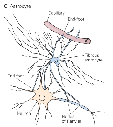
Principles of Neural Science - McGraw Hill (2021): Part 2 - Chapter 7- while not part of the BBB, they do help Ependymal cells
- modulate the milineu of the CSF
- Can take up potassium ions
- Regulates GABA and inactivated neurotransmitters
Microglia Cells
Generally thought of as the brains immune cells, are the first line of defense in the brain and spinal cord. While they can adapt to various phenotypes depending on their location and situation, they do have the following general tasks:
- Remove plaques, dead cells
- Remove Unnecessary neurons and synapses
- while this is especially true during brain development, they do still maintain this function afterwards. This is an active area of research!
- Destroy infectious agents
Goldman-Hodgkin-Katz (GHK) equation
Used to calculate the membrane potential, given the interactions of the main ions involved with neuron membranes (Potassium, Sodium, and Chlorine.
- Vm is the membrane potential. This equation is used to determine the resting membrane potential in real cells, in which K+, Na+, and Cl- are the major contributors to the membrane potential. Note that the unit of Vm is the Volt. However, the membrane potential is typically reported in millivolts (mV). If the channels for a given ion (Na+, K+, or Cl-) are closed, then the corresponding relative permeability values can be set to zero. For example, if all Na+ channels are closed, pNa = 0.
- R is the universal gas constant (8.314 J.K-1.mol-1).
- T is the temperature in Kelvin (K = °C + 273.15).
- F is the Faraday's constant (96485 C.mol-1).
- pK is the membrane permeability for K+. Normally, permeability values are reported as relative permeabilities with pK having the reference value of one (because in most cells at rest pK is larger than pNa and pCl). For a typical neuron at rest, pK : pNa : pCl = 1 : 0.05 : 0.45. Note that because relative permeability values are reported, permeability values are unitless.
- pNa is the relative membrane permeability for Na+.
- pCl is the relative membrane permeability for Cl-.
- [K+]o is the concentration of K+ in the extracellular fluid. Note that the concentration units for all the ions must match.
- [K+]i is the concentration of K+ in the intracellular fluid. Note that the concentration units for all the ions must match.
- [Na+]o is the concentration of Na+ in the extracellular fluid. Note that the concentration units for all the ions must match.
- [Na+]i is the concentration of Na+ in the intracellular fluid. Note that the concentration units for all the ions must match.
- [Cl-]o is the concentration of Cl- in the extracellular fluid. Note that the concentration units for all the ions must match.
- [Cl-]i is the concentration of Cl- in the intracellular fluid. Note that the concentration units for all the ions must match.
Ion Channels
Structures embedded in the phospholipid bilayer to allow charged ions to pass through, though often with some form of control, and falls under various classes, including (but not limited to):
- Random: known as leak / non-gated channels. Essentially no active control of having ions pass through
- Membrane Potential: Also known as voltage gated channels, can be toggled with changes to voltage across the membrane
- Ligand Gated Channels: Toggles channel based on the availability / absence of a chemical messenger
Kinetics
The probabilistic temporal dynamics of a molecular reaction from one state to another.
Example: Gated channels with fast kinetics can toggle faster
Length Constant
The distance it takes for a potential charge to fall off to about 36% of its initial value along the neuron membrane
Example problem
There’s a voltage change of 100 mV at point A. Point B is 2 mm away from point A, and point C is 6 mm away from point A. The length constant for this neuron is 2 mm.
Since B is 2 mm away from A, which is equal to one length constant, we know that the voltage change will decrease to about 37% (of the original change). So, at point B, the voltage change will be (100 mv)(0.37) = 37 mV.
C is 6 mm away from A, which is 3 length constants away. We can use the same logic and find that the voltage change at point C is (100 mv)(0.37)3 = (100 mv)(0.37)(0.37)(0.37) = ~5 mV.
So the length constant tells us information about how far voltage changes can travel in a neuron before attenuating to very small values. The 37% approximation comes from a model of exponential decay.
Milieu
Another term for "Environment", as in cellular environment / cellular Milieu
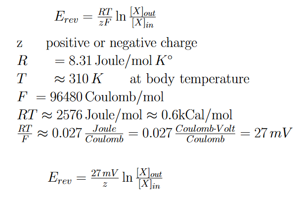
Nerst Equation
In the context of Neuroscience, this can be used to define the electrochemical equilibrium for a single particular ion across the cellular membrane. Can be used to find the Reversal Potential (also called the Nerst Potential), which is the electrical potential that balances against the concentration gradient.
Neuron
The basic nervous system cell responsible for receiving, processing, and outputting signals due to being electrically and chemically excitable and due to their asymmetric makeup.
Neurons are not the primary type of cell of the nervous system, they are outnumbered by Glial cells three to one!
(Principles of Neural Science - McGraw Hill (2021): Part 1 - Chapter 3)
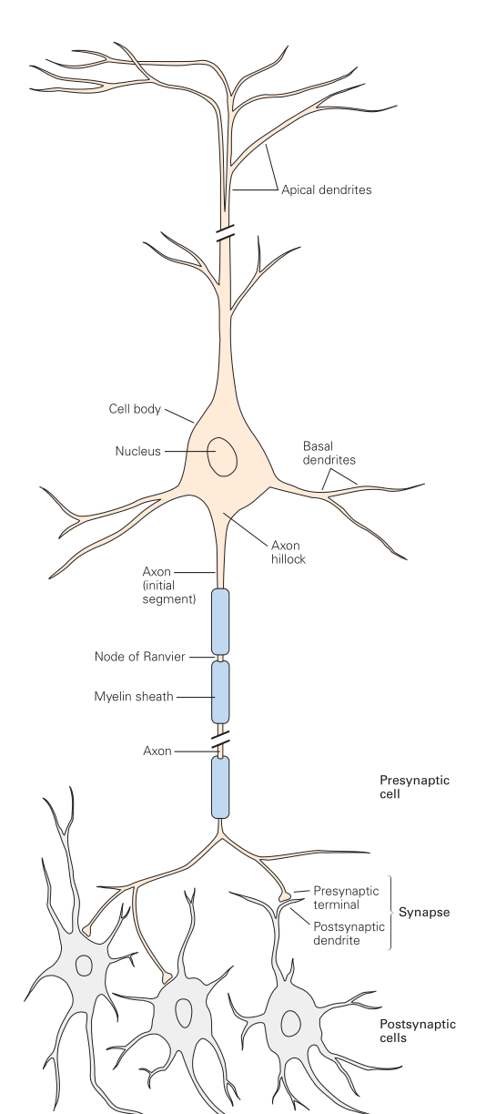
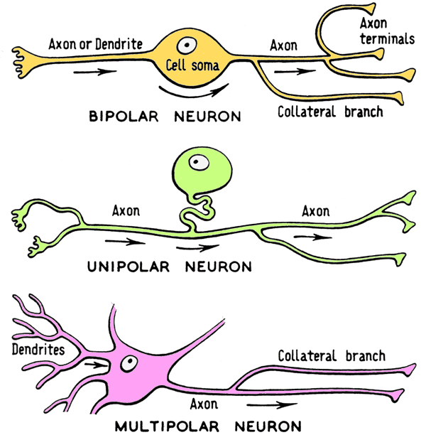
The basic structure of a neuron consists of:
- Dendrites: Branches coming off the center cell body, are generally small but numerous, and act as the input for the neuron
- Cell body / Cell Soma: The 'Center' of the neuron, which is where the nucleus is contained
- Axon Hillock: The protrusion of the soma before it becomes the axon, where all input signals from the dendrites are combined where they may reach the firing threshold and trigger an action potential
- Axon: The output of the neuron, where action potentials flow out to other enervated neurons or receptor cells
There are hundreds of neuron types and they can be categorized in various manners:
There are hundreds of neuron types and they can be categorized in various manners:
Polarity
Neurons can be structured in slightly various manners to better fit their specific role. They can be Bipolar, Unipolar, or Multipolar.
Afferent / Interneuron / Efferent
Names of categories referring to the general functional role of a neuron.
- Sensory / Afferent: Receives data from the outside world.
- Tend to be unipolar or bipolar.
- Example: Photo-receptor.
- Interneuron: Neurons that connect to other neurons. Does not receive input from the world, nor directly control any response. Are typically involved in information processing.
- Tend to be multipolar.
- Example: Most of the neurons in the brain.
- Motor / Efferent: Outputs a response to the outside world. Typically to a muscle but potentially also to things like glands.
- Tend to be multipolar.
- Cell body is often found in the spine
- Example: Nerve innervating a muscle
Node of Ranvier
Gaps between individual myelin sheaths along a neuron. Critical for Saltatory Conduction
Ohm's Law
States that Current = Voltage / Resistance
Peripheral Nervous System (PNS)
A division of the nervous system, composed of nerves that branch out from the CNS (like out of the spinal cord). Basically anything part of the nervous system that isn't the Brain or Spinal Cord themselves.
Permeability
The ease at which a particle can pass through the membrane
Phospholipid Bilayer
The structure of most cell membranes, include of Neurons.
- Is difficult for charged ions to pass through directly
- They must use (often controllable) channels
- Small non-polar molecules like CO2 and O2 can pass directly through the bilayer, larger ones sometimes can slowly
-
- some polar molecules can slowly pass through, like water
- Water usually passed through Aquaporin Pores
- some polar molecules can slowly pass through, like water
- Larger molecules often require the aid of structures to pass through
-
The Phospholipid Bilayer is composed primarily of Phospholipid molecules.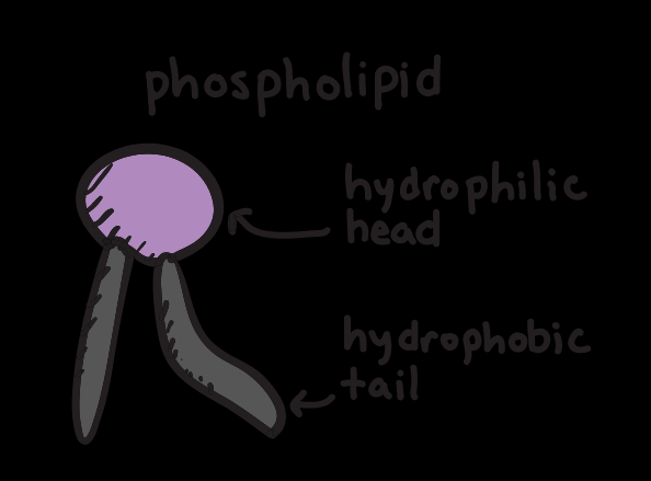
The bilayer is composed of two layers of these phospholipids, with the hydrophilic head facing the outside / inside environments and the hydrophobic fatty acid tail being within the layer sheet.
- This makes sense, since the hydrophilic head prefers aqueous environments, this is a self organizing structure!
- The centered hydrophobic tails make it difficult for water to pass through the bilayer directly
- Cholesterol is also a big component of the cell membrane. It accomplishes several tasks, such as maintaining membrane "fluidity" across a wide temperature range, and also can help as a glue keeping phospholipids together.
Refactory Period
A period of time where we can't (easily) stimulate another action potential in a neuron.
Two Types:
- Absolute Refactory Period: caused by inactivation kinetics of sodium channels. While the sodium channels are in that state, we cannot initiate another action potential AT ALL.
- Relative Refractory Period: Is during the tail end of the action potential, when the sodium voltage gated channels are active but the potassium channels are open still. We can drive another action potential with a strong enough stimulus since the membrane is starting below the resting potential
Resistance
The measurement of resistance in current, measured in ohm (Ω). Inverse of Conductance (which is measured in Siemens, or S or simply 1/Ω). Ion Channels have resistance, and increasing the permeability and channel count decrease resistance. In addition, the saltier the cytoplasm, the lower the electrical resistance,
Saltatory Conduction
When during an action potential, voltage "jumps" from one Node of Ranviers to the next (running within the nerve fibers wrapped in myelin sheath,. The voltage strength drops during the process, which is why it needs to be 'boosted' and the Node of Ranviers.
Voltage / Potential
Voltage is a measure of 'pressure' of the electrical force from one area to another.
Examples:
- Batteries use chemistry to create a voltage differential between 2 ends of the battery cell
- Neurons use ion gradients (not chemistry!) across the cell membrane
In neurons, when measuring the voltage, we always use the outside of the cell as ground measurement
'Potential' is often used as a synonym for Voltage
There are some potentials to know about:
- Resting Potential: The stable membrane potential when a neuron is at rest (IE not firing)
- Action Potential: The series of ion concentration changes that change the voltage across the membrane as a neuron is firing
- Breakdown Potential: An extreme case, the point at which a potential is so strong that ions simply punch straight through the membrane

