Ch 2: Advantages and Limitations of Time- and Time-Frequency-Domain Analysis
This section is a WIP!
Ch 1: The Purpose of This Book, Who Should Read it, and How to Use it
This chapter is mainly an introduction for the book, discussing that it serves as an overview of the analysis that comes with EEG (but it can be done with other time-varying signals), but it is not exhaustive. Skipping over most of it here as it is not relevant to this summary
1.1 Cognitive Electrophysiology
The field of study known as Cognitive Electrophysiology can be thought of as a spectrum. One side is Cognitive psychology, which concerns itself with the cognitive components of the brain. The other side is Electrophysiology, which concerns itself with the functional properties of the neural networks that make up the brain.
Ch 2: Advantages and Limitations of Time- and Time-Frequency-Domain Analysis
This chapter discusses the different categories of analysis and their pros/cons, some discussion of EEG vs MEG, brain network scales, and brain localization
2.1 Why EEG?
EEG has a high temporal resolution (unlike fMRI), which allows it to capture the cognitive events that occur within in 100s of milliseconds, from activity in the 4-8 hz theta-band oscillations all the way to 30-80 hz gamma band. EEGs also directly measure neuronal activity of populations of neurons, which is very prominent when measuring oscillations. EEG is also multidimensional, as the data compromises information across time, space / location, frequency, and power / strength.
2.2 Why Not EEG?
EEG is not suited when looking for precise locations, and (generally) isn't suited for deep brain structures. IE it is a bad tool for finding " MEG may be better suited if these are you concerns.
EEG is likely to be a suboptimal method for any research questions involving "where in the brain does process X occur or is information Y stored"
EEG / MEG are both unsuited if trials of the experiment last longer than a few seconds or if time is highly variable. This makes it poorly suited for complex cognitive task studies, or those concerning social and emotional responses. The lower temporal resolution of fMRI makes it a better fit instead.
2.3 Interpreting Voltage Values from the EEG Signal
Measurement of EEG signals is typically microvolts, while with MEG it is femtotesla. For EEG, these absolute microvolt values arent helpful, as they can differ in magnitude due to irrelevant factors such as skull thickness or shampoo used or dipole orientation (similar irrelevant factors exist for MEG). Transformations such as decibel normalization may be preferred instead as then there can be comparisons made between trials.
2.4 Advantages of Event-Related-Potentials
2.5 Limitations of ERPs
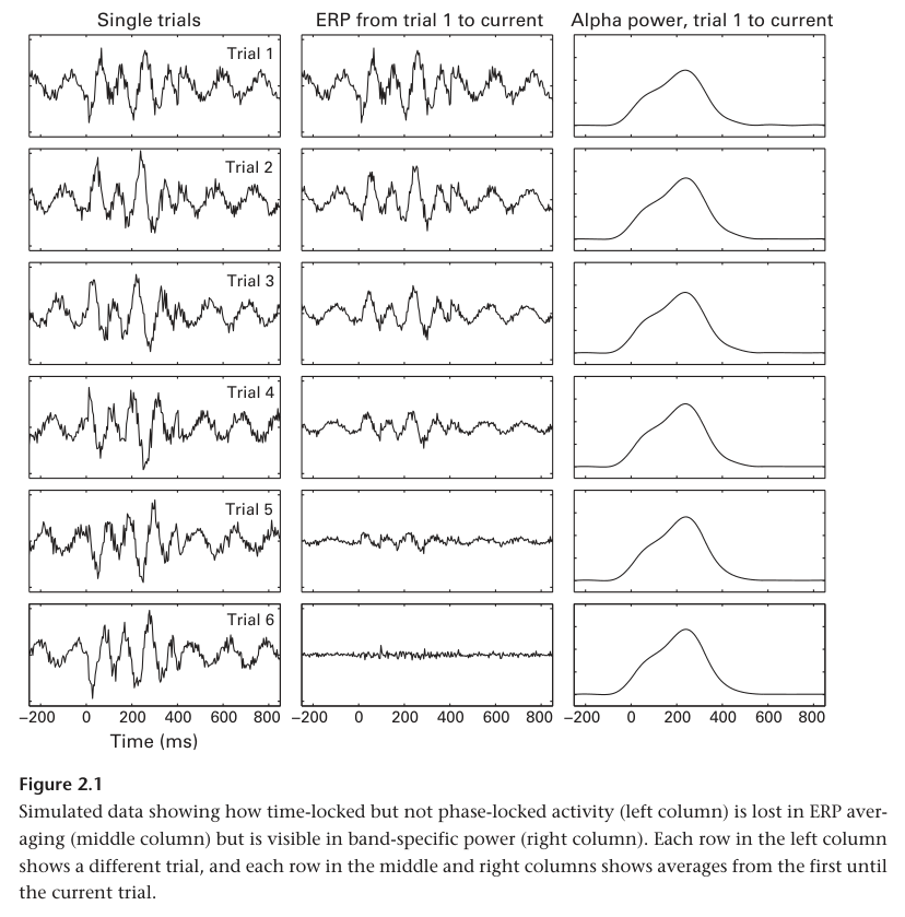
ERPs actually reveal rather little data from EEG signal, which makes it difficult to interpret null results (when your experiment finds nothing). ERPs also cannot be averaged with other trials, as it reduces the output to noise. It is also harder to link to physiological mechanisms since the neurophysiological mechanisms that produce ERPs are less understood than those that produce, for example, oscillations.
2.6 Advantages of Time-Frequency-Based Approaches
This is a diverse set of analysis. Generally, it will be easier to use these to interpret results in terms of neural oscillations, which is important since oscillations are a fundamental neural mechanism and studied well. They show a wider set of information capturable from EEG vs just ERPs, and thus can be used to answer more questions.
2.7 Limitations of Time-Frequency-Based-Approaches
As a result of time-frequency decomposition, temporal resolution will be lost. In general, temporal resolution will be worse than what is achievable with fMRI, but worse than with ERP analysis (theoretically, low pass filtering diminished its temporal resolution). And while not a limitation strictly speaking, Time-Frequency analysis is a very wide category, and it may be easy to get lost on the specific analysis to do.
2.8 Temporal Resolution, Precision, and Accuracy of EEG
Terminology is important. Temporal Resolution refers to the number of data samples per unit time, while precision refers to how accurate those sample are with the timing of the actual biophysical events in the brain that produced the EEG signal. Temporal resolution is initially determined by the sampling rate of the EEG headset, which is usually within a few hundred to a few thousand Hz (generally between 250 - 1000 Hz). Precision depends on the analysis. Unfiltered ERPs tend to have the best precision. However, precision drops with things like sliding window type filters causing temporal leakage of information. For these reasons, while time-frequency analysis requires high temporal resolution input, its output loses enough precision that it can also be downsampled to a lower temporal resolution without any meaningful loss of context (say to 50Hz).
2.9 Spatial Resolution, Precision, and Accuracy of EEG
Spatial Resolution refers to how close a signal detected can be placed to its actual source origin. EEG's spatial resolution depends on the number of electrodes (generally starting at 32, up to several hundred), but EEG is generally considered to have low spatial resolution, accuracy, and precision. The precision can be 'improved' via spatial filters such as surface Laplacians. However, spatial accuracy can remain low, as activity recorded from an electrode can come from various brain regions close and distant to the electrode depending on various factors. Generally though, EEGs can best localize signals to relatively large cortex regions of several cubic centimeters.
2.10 Topographical Localization versus Brain Localization
Topographical Localization refers to identifying the electrodes that show the max effect of a signal. This should not be mistaken for Brain Localization, as we do not technically know if the neurons responsible for the signal were directly under that electrode.
2.11 EEG or MEG?
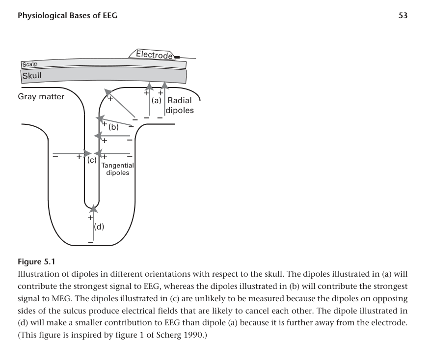
EEG and MEG measure similar physiological properties, but the results may not be the same. This is because EEG is maximally sensitive to radial sources on the gyral crowns (due to the direction of the electric field), and MEG is sensitive to tangential sources.
Furthermore, MEG is better suited to detecting higher frequency activity (above 60 Hz).
In more practical differences, EEG is portable (MEG in a special environment) but MEG doesn't require any special gels (to be fair some newer EEGs don't require gel either). MEG source reconstruction (localization) tends to also be a bit more accurate simply because most EEG setups do not have solidly fixed electrode locations, unlike MEG.
2.12 Costs of EEG Research
This section points out that while EEG seems cheaper initially compared to fMRI or MEG, replacing consumable parts gets expensive quickly, often to the point that prices of these tools become similar per subject by the end anyways. Ergo, its best to focus on what tool best answers the hypothesis, not what seems cheapest.