Ch 5: Introduction to the Physiological Bases of EEG
This chapter discusses the mechanisms to how neuronal activities end up as signal data recorded by EEG (and MEG)
The author states this is merely a brief summary, and recommends the papers here (free), here, here, and here (free).
5.1 Biophysical Events That Are Measurable with EEG
When Neurotransmitters activate ion channels in neuron membranes, ions flow through the membrane causing a change a potential, resulting in an electrical field. While the field of one neuron is too weak for EEG, the fields of 100s-10,000s of neurons in sync can be detected by EEG electrodes. Ergo, EEG is the summation of excitatory and inhibitory potentials of ensembles of neurons with parallel geometric orientation
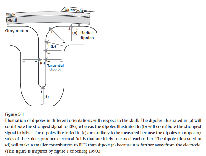
- Note that it is important that they are parallel because if each neuron was pointed in a random direction, their fields would cancel each other out on average.
Distance to electrodes does matter, which is why EEG signal is chiefly dominated by neurons in the outer cortical layers (between 10,000 to 50,000 neurons).
- Since electrical conductivity through air is nearly non-existent, gels, and other substrates must be used by EEG electrodes to make contact with the scalp. MEG does not have this limitation / requirement as magnetic fields are not impeded by air
Like all brain Imagine techniques, EEG cannot measure everything. Along side not being able to measure individual neurons, it cannot segregate between various neurotransmitters / neuromodulators, it also cannot effectively measure electrical activity (even strong) from populations on opposite sides of a sulcus (brain fold) as the electric field would cancel itself out overall.
EEG generally has a very difficult time measuring deep brain structures. The only way to do it is if the field is very strong, and you need thousands of trials to filter the data from the noise. The reason for this difficulty is that the region is physically far from the electrode, and deeper populations of neurons tend not to be structured in parallel orientations. This is why imaging techniques such as fMRI is better suited for these tasks.
Very slow oscillations (under 0.1 Hz) can be difficult to measure in EEGs due to hardware limitations with amplifiers and filters often employed. Signals that are high frequency (above 100 Hz) are difficult to measure instead due to the simple face that higher frequency oscillations have lower power and easily get lost in the noise. MEG may be a better fit for this frequency range.