Ch 5: Introduction to the Physiological Bases of EEG
This chapter discusses the mechanisms to how neuronal activities end up as signal data recorded by EEG (and MEG)
The author states that this is merely a brief summary, and recommends the papers here (free), here, here, and here (free).
5.1 Biophysical Events That Are Measurable with EEG
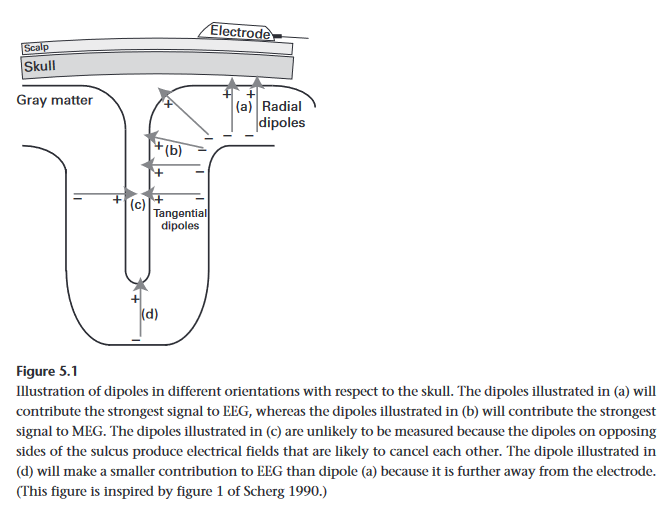
When Neurotransmitters activate ion channels in neuron membranes, ions flow through the membrane causing a change a potential, resulting in an electrical field. While the field of one neuron is too weak for EEG, the fields of 100s-10,000s of neurons in sync can be detected by EEG electrodes. Ergo, EEG is the summation of excitatory and inhibitory potentials of ensembles of neurons with parallel geometric orientation
- Note that it is important that they are parallel because if each neuron was pointed in a random direction, their fields would cancel each other out on average.
Distance to electrodes does matter, which is why EEG signal is chiefly dominated by neurons in the outer cortical layers (between 10,000 to 50,000 neurons).
- Since electrical conductivity through air is nearly non-existent, gels, and other substrates must be used by EEG electrodes to make contact with the scalp. MEG does not have this limitation / requirement as magnetic fields are not impeded by air
Like all brain Imagine techniques, EEG cannot measure everything. Along side not being able to measure individual neurons, it cannot segregate between various neurotransmitters / neuromodulators, it also cannot effectively measure electrical activity (even strong) from populations on opposite sides of a sulcus (brain fold) as the electric field would cancel itself out overall.
EEG generally has a very difficult time measuring deep brain structures. The only way to do it is if the field is very strong, and you need thousands of trials to filter the data from the noise. The reason for this difficulty is that the region is physically far from the electrode, and deeper populations of neurons tend not to be structured in parallel orientations. This is why imaging techniques such as fMRI is better suited for these tasks.
Very slow oscillations (under 0.1 Hz) can be difficult to measure in EEGs due to hardware limitations with amplifiers and filters often employed. Signals that are high frequency (above 100 Hz) are difficult to measure instead due to the simple face that higher frequency oscillations have lower power and easily get lost in the noise. MEG may be a better fit for this frequency range.
5.2 Neurobiological Mechanisms of Oscillations
In this context, oscillations refers to the alterations between state of excitability of neurons. THere are several mechanisms for these oscillations. Two major examples include:
- GABA inhibitory neurons and Pyramidal excitatory neurons
- pyramidal cells become activated, they excite each other as a closed feedback loop
- GABA inter-neurons in the network also get activated, start sending inhibition signals to the population
- signal drops as the the pyramidal cells become inhibited, causing the GABA neurons to cease reviving the signals to fire
- pyramidal cells, no longer receiving inhibitory signals, start firing excitatory signals again
- repeat
- In other cases, some purely excitatory networks and purely inhibitory networks are coupled together, and interact in a similar fashion
5.3 Phase-Locked, Time-Locked, Task-Related
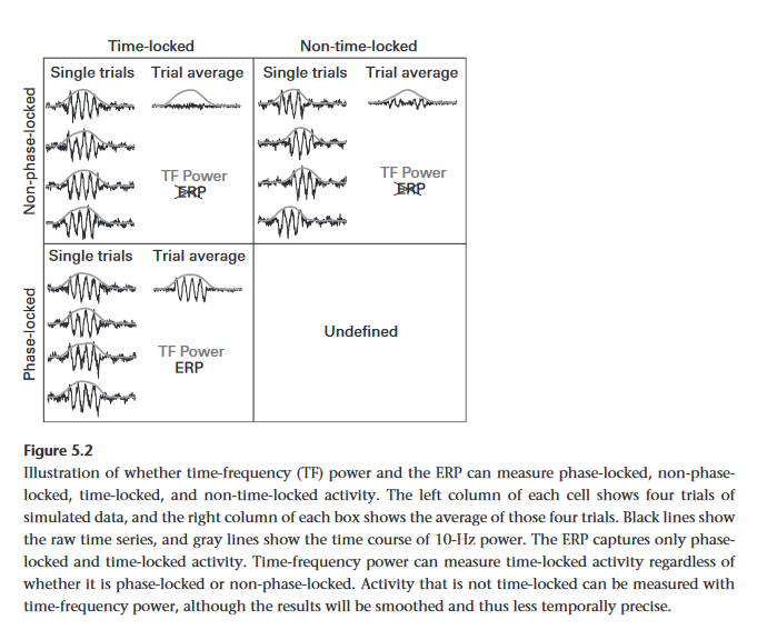
Phase locked (evoked) activity is phase-aligned with the time = 0 event. The benefits of this is such activity can be observed through time-domain averaging (ERPs) and time-frequency-domain averaging.
Non-phase locked (induced) activity is not phase aligned with time - 0 events. While this activity can be observed in time-frequency0dinaub averaging, it cannot be observed in time-domain averaging (ERPs).
Currently, its unclear why some responses are phase locked and others not.
Background activity does not change in response to task events. It generally adds noise to data in an experiment, so often baseline normalization is used to remove it.
5.4 Neurophysiological Mechanisms of ERPs
There are several theories on the functioning mechanisms behind ERPs and why they occur
- Additive
- Event causes a signal "spike" that is added to background oscillations. Background oscillations can be averaged away to show only the ERP
- Phase Reset
- Event causes several oscillations to reset in phase, causing them to initially accumulate together in a spike of activity before they lose coherence again
- Amplitude asymmetry or baseline shift
- While electric currents from neurons are polarity balanced, outward currents may be less detectable from the scalp. This asymmetry causes the oscillations measured by EEG. Changes in overall power can alter this asymmetry
There is still a lot of active debate on the theories involved, as the actual mechanisms of ERPs still needs further research.
5.5 Are Electrical Fields Causally Involved in Cognition?
Technically its unclear, but there is some evidence to support this:
- Hebbian learning in the hippocampus occurs at 40 Hz oscillation
- Optogenitics - genetically modified mice have neurons that respond to certain wavelengths, so we can control their oscillations with light. Experiments with this showed that oscillations were important for problem solving
- Studies show that transformation of information across regions works most efficiently where regions are phase synchronized
- Transcranial Alternating Current Stimulation (TACS) uses 2 electrodes on the scalp to pass through a alternating current from 0.1 to 100 Hz. It is show that various frequencies of stimulation affected certain functions
5.6 What if Electrical Fields Are Not Causally Involved in Cognition?
Its possible the work on the role of neural oscillations is wrong, and theories based on them as well. It does not mean this field of study is to be lost, since electrical fields produced by neuron populations is still a powerful insight of brain function.
In the same vein (heh), Blood Oxygenation Level Dependent (BOLD) fMRI imaging scans for oxygen saturation in various areas of the brain over a period of time to ascertain activity in various regions. No one states that cognition is a direct result of changing oxygen levels in different parts of the brain, but BOLD still remains a powerful tool in neuroscience research


